
Ultrasound of the pelvis for diagnosing fibroids
The womb and ovaries are located in the lower part of the abdomen. There are many pathologies of the womb the most common one is fibroids. Approximately 80% of people with a womb will develop fibroids at some point in their life. Fibroids are non-cancerous / benign growths that develop in or around the womb (uterus).
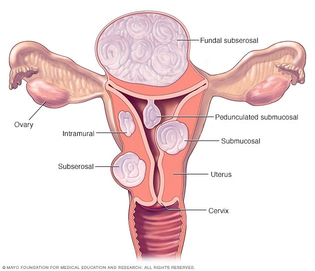
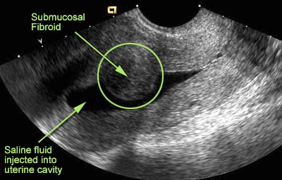
Types of fibroids
Symptoms
These include; heavy menstrual bleeding, long menstrual periods, bleeding in between periods, pelvic pressure or pain, frequent urination, difficulty emptying the bladder, constipation and backache or leg pains.
Preparation and Ultrasound examination
Your Doctor will request a pelvic or gynaecology scan to be caried out. Two scans are likely to be formed with your consent; a transabdominal scan and a transvaginal scan. Both scans will assess the womb, the lining of the womb, the ovaries and areas adjacent to the ovaries.
Transabdominal scan requires a full bladder that is 2 cups (500mls) of water an hour before the scan. The full bladder will act as a window to view the pelvic organs. You will be required to lie on your back. Ultrasound gel is applied onto the pelvic area. The ultrasound probe or camera is moved across the pelvis whilst images will be recorded. This scan is usually less than 10 minutes.
Transvaginal scan requires an empty bladder. You will be required to undress from the waist down and given towels to cover yourself. You will be asked to lie on your back and asked to bring your knees apart for the insertion of the ultrasound probe / camera. The ultrasound probe is sterilised before the scan and is covered with a non- latex or a latex cover. Ultrasound gel is applied at the tip of the camera for lubrication and also for clear visualisation of the organs. The procedure should not be painful but unusual. This scan is usually less than 10 minutes.
After the scan the gel is wiped off. The Sonography practitioner will report findings to the referring Doctor. The referring Doctor will advise you if there is any specialist referral needed.


There are no comments yet, why not be the first